Symbiosis: Entoprocta (comensal)


Ophiuroidea – Entoprocta
  • Ophiuroidea: Ophioplinthus gelida: host 
  • Entoprocta: Loxosomella sp : commensal

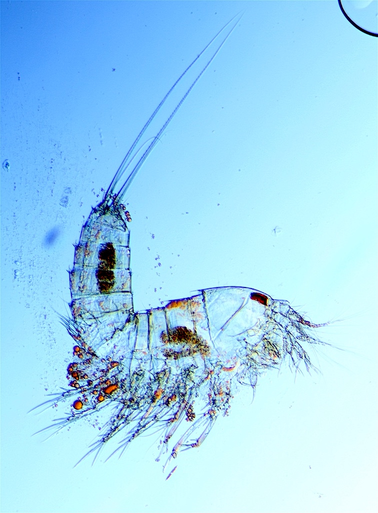
Loxosomella sp. as commensal of Ophioplinthus gelida (Koehler 1900), from which it obtains a substrate that provides its mobility to the food sources, the advantage of not being buried by the sediment as well as certain protection from predators Emschermann (1993). For Ophioplinthus it seems to be no benefits at all.


Loxosomella is mainly located at the edges of dorsal interradii as well as on the lateral plates along the arms, being able to appear with a high density of individuals. They can also appear on the ventral part of the disk, but in a scarce proportion.
Entoprocta (Kamptozoa): colonial or solitary suspensivore lophophorates whith the anus inside of the tentacle crown (lophophore), unlike the bryozoans (Ectoprocta) Loxosomella are solitary entoprocts.

1. Extended lophophorate   2. Bud    3. Contracted lophophorate

Materials studied from Expedition ANTARKTIS- XXlll / 8 Polarstern 2006/2007  in  Joinville Island (3 brittle stars with Loxosomella). They correspond to the same geographical area that the specimens studied by Emschermann to Loxosomella antarctica (Weddell Sea and Bransfield strait).
Specimen conserved in 70% ethanol, all  photographs were made in ethanol.
Collected by Pablo J. González-López.
Identified by Rafael Martín-Ledo.
The study was made using Motic SMZ-168 TL stereo microscope.

References

Emschermann (1993) On antarctic Entoprocta: Nematocyst-like organs in a loxosomatid, adaptive developmental strategies, host specificity, and bipolar occurrence of species. Biol. Bull 184: 153-185

July 2010

Marine Aquarium -British Antarctic Survey (BAS)-


One of the most impressive experiences of this summer was that of having live Antarctic brittle stars in my hands – and I took that unique opportunity to identify them. Where? In the Marine Aquarium of British Antarctic Survey (Cambridge).

Different aquariums with various zoological groups carefully selected according to their way of life: cnidarians, teleosts, pycnogonids or echinoderms live in marine aquariums situated in a cold room, specially designed to shelter the animals at Antarctic waters temperature.


Sandy Cordiner-Lawrie, Marine Aquarium Manager of Biological Sciences Division (BAS), combining professionalism and affection takes daily care of the fragile marine creatures caught selectively by scuba-divers in Rothera Research Station (Adelaide Island, Antarctida).Among her numerous responsibilities there are those of control of the physico-chemical parameters of the water and maintaining it clean, feeding the animals etc.


Different studies are being carried out, from metabolic routes to growth physiology, and the isolation of the specimens is often needed.


There was a really interesting phenomenon – an “echinoderm swarm”, some kind of caotic concentration of the animals on the wall of one of the tanks, while the rest of the tank was almost empty. It seemed that sea urchins, starfishes (the predator Labidiaster !) and sea cucumbers were desperately seeking a refuge among themselves.


Thank you, Sandy!


September 2010

Historia de un crimen


Luria lurida (Linnaeus, 1758) es un molusco gasterópodo de la familia de las cypraeas que vive en aguas poco profundas. 
Una luria volteada, se distinguen el manto recubriendo la concha, el pié con restos de sedimentos por donde se había desplazado, un ojo y los tentáculos.

Por el día permanece metida en su concha, oculta entre las rocas y, por la noche sale para alimentarse de esponjas, cnidarios o pequeños crustáceos. Es entonces cuando el manto* del cuerpo del animal envuelve la concha y la frota continuamente dándole el típico brillo de las cypraeas.


Un cangrejo ermitaño (Clibanarius aequabilis), se asoma en una Luria.

Cuando se encuentra una cypraea, en la línea de varado de cualquier playa, suele haber perdido su brillo. Al morir el animal la concha servirá de hogar a un cangrejo ermitaño y puede que se cubra de organismos epizoicos*, o simplemente rodará por acción de las corrientes durante días o semanas antes de ser depositada en la playa.


Ejemplar recolectado en la playa de Las Américas (Tenerife).

Así pues, encontrar un ejemplar casi intacto nos indica que el animal murió hace muy poco tiempo, quizás horas. 
Buceando en la La Caleta de Adeje (Tenerife), tuve la suerte de encontrar este ejemplar.


Ya en casa, me di cuenta que el animal no había muerto "de viejo". En la concha se puede ver un orificio muy pequeño. Es la prueba del delito; hacía muy pocas horas que se había cometido un crimen. 
El orificio es la abertura practicada por un pulpo, empleando su rádula, para poder introducir su jugo gástrico que contiene una cefalotoxina. Esta sustancia causa la distensión de la musculatura del animal, el cual al relajarse es fácilmente sacado de su concha, por la acción prensil de los tentáculos del pulpo y, ya está listo para ser devorado.

Orificio en la concha de una luria realizado por un pulpo.

Una luria, izquierda,  su manto recubre la concha que de tanto frotarla quedará brillante. Un pulpo, derecha, agazapado en una poza de la rasa en bajamar.

Esta relación interespecífica entre pulpos y lúridas la podemos encontrar en fósiles del pleistoceno (hace 100.000 años). El ejemplar de la siguiente foto lo encontré en el yacimiento paleontológico El Guincho-El Bunker, en Playa de las Américas. Se puede apreciar el orificio realizado por el pulpo y que supone la evidencia de la predación.


Manto: parte dorsal del cuerpo de un molusco; recubre las vísceras y forma la concha; está constituido por músculos y forma el pie que utiliza el animal para su movimiento.
Epizoico: hace referencia a organismos que viven sobre animales, como es el caso de algas sobre conchas de moluscos.
Rádula: estructura "dentaria" de los moluscos empleada para raspar el alimento.

Fuentes
Wodinsky J (1969) Penetration of the shell and feeding on gastropods by octopus. American Society of Zoologists, 9

Symbiosis: Amphiura - Eulimidae (parasitism)

Amphiura joubini: host
Eulimids: endoparasite


In a Amphiura belgicae specimen, caught in South Georgia, Mortensen (1936) described a gastropod that could belong to the same species as the one from the present study. Mortensen described as a possible mechanism of parasite infestation its entrance through an orifice. He also indicated that the adult looked like a sack and had no visible shell.

In the examined specimens of Amphiura joubini from South Shetland we can  see externally one or two large whitish interradios. When we opened the animals, we saw a bag with numerous white or flesh colored shells inside the coelom, the adult being globular.


There is no observable orifice through which the gastropod entered, so there is a possibility of its regeneration. However, in one of the specimens that had two parasites in contiguous interradios, the other interradios are open ventrally, which leads us to think that this would be an “exit” mechanism for the gastropods.
This parasitism does not necessarily mean the death of the brittle star, possibly due to its great regenerative capacity. Although this is only a supposition, based on the large number of examined specimens of Amphiura joubini with badly damaged interradios due to the process of youth parasitic forms leaving their host.
There have also been numerous open specimens of Ophiolimna antarctica, without most of the aboral disk, it is hard to find a “whole” specimen in our collection. In one of the specimens a pair of juvenile gastropods were found, so it does not seem to be a specific Amphiura genus parasitism.
Although there is a reference in the literature on the presence of internal eulimidae parasites, not yet been identified at specific level (Anders Waren, pers. Comm.)


Live Amphiura jouvini

Symbiosis: Cancerilla - Ophioika (parasitism)


Ophiuroidea – Copepoda

First the ectoparasites copepods of Antarctic ofiuroids were cited by Mortensen (1936), but without identifying the species, it was Heegaard (1951) who identified them. In the taxonomic literature they have also been cited by Madsen (1967).

Ophiacantha antarctica: host
Cancerilla sp: ectoparasite


The copepods are adhered to the dorsal surface of the disc. Females are carrying their bags of eggs like balls, males have not been found.

Ophiacantha antarctica: host
Ophioika sp: endoparasite


Copepods with modified morphology occupy nearly an entire interradius of the disk. In this species very small males are situated inside of females brood-pouch.
In his review of Ophiuroidea reproduction, Hendler (1991), cites a work of Bartsch (1975) in which he described a castration as a consequence of an ectoparasite copepod (Cancerilla oblonga) in Amphiura capensis. This is an issue to investigate whether in Antarctic ofiuroids there are similar consequences.

Specimens conserved in 70% ethanol, all  photographs were made in ethanol.
Collected by Pablo J. González-López. Cruiser ANTARKTIS XXIII/8  Polarstern.
Identified by Rafael Martín-Ledo.
The study was made using Motic SMZ-168 TL stereo microscope.

References

Heegaard, P (1951) Antarctic parasitic copepods and an ascothoracid cirriped from brittle-stars. Vidensk. Medd. Dan. Naturhist. Foren. 113, 171-190

Hendler, G (1991) Echinodermata: Ophiuroidea. In: Giese, A.C., 

Pearse, J.S. & Pearse, V.B. (Eds.), Reproduction of marine invertebrates: echinoderms and lophophorates. The Boxwood Press, Pacific Grove, CA, pp. 355–511

Madsen FJ (1967) Ophiuroidea. B.A.N.Z. Antarctic Research Expedition (1929–1931) under the Command of Sir Douglas Mawson Rep Ser B 9:123–145

Mortensen T (1936) Echinoidea and Ophiuroidea. Discovery Reports, National Institute of Oceanography Cambridge 12:199–348

July 2010

Benthic Copepod (Harpacticoida)

Samples from Bay of Santander 

The benthic copepods live in the ocean floor sand and on algae.
Features of Order Harpacticoida:
Anterior part of body (metasome), not conspicuously distinct from the posterior part (urosome). First antenna shorter than the cephalic segment. Females frequently have one median egg sac.


Plan Fluor 10x                                                               Canthocamptus sp

Plan Fluor 10x                                                      Canthocamptus sp

Plan Apo 20x                                                         Canthocamptus sp

Plan Fluor 10x                                        Canthocamptus sp

Plan Apo 20x                                       Canthocamptus sp

Plan Apo 20x                                                         Canthocamptus sp

     
Plan Fluor 40x

Plan Fluor 10x 

 Plan Fluor 10x 

Plan Apo 20x

Plan Fluor 10x                                                      Porcellidium sp


Nikon Eclipse E600 microscope / Pol - DIC 
Canon EOS 80D camera

Symbiosis: Ophioplinthus - Ascothorax (parasitism)

Ophioplinthus brevirima: Host
Ascothorax sp: endoparasite


Inside there were several parasites (Ascothorax sp) with globose aspect; the smaller ones (probably males) with better distinguished morphology having two valve, while the larger specimens are possibly females. This difference in size, and better conservation of structures in male specimens is a common phenomenon in many parasitic crustaceans.

Ophioplinthus brevirima has short genital  slits, hence its specific epithet. In the studied specimens one of the slits, which coincides with the deformed area of the disc has a deformation making it larger, and the proximate plates look abnormal. It is likely had been caused when the endoparasites were leaving their host.


This is supposedly the first record of endoparasites for Ophioplinthus brevirima.

Materials studied from Expediton ANTARKTIS-XXIII/8 Polarstern 2006/2007 in Snow Hill and Dundee Islands. Specimens conserved in 70% ethanol, all photography were made in ethanol using Motic SMZ-168 TL  stereo microscope.

Collected by Pablo J. González-López
Identified by Rafael Martín-Ledo
July 2010


Live Ophioplinthus brevirima, covered by Iophon sponge


Fósiles con simetría bilateral

Simetría bilateral: un solo plano, el sagital, puede dividir el cuerpo del animal en dos partes simétricas (mitad derecha e izquierda).

Puedes hacer click sobre las imágenes para verlas a un tamaño mayor.


Fuentes y materiales
- EDUCTRADE (1990). Colección de fósiles y guía de utilización.
- Colección de fósiles del IES Leonardo Torres Quevedo (Santander).